Project Details
- International speaker : Shinji Deguchi, Osaka University, Japan.
- In-house speaker : Claudia Prieto, P. Catholic University of Chile, Chile.
- Generous prizes : Three (3) best oral contributions, three (3) best posters!
- Location : Aula Lassen AL4, Campus San Joaquin P. Universidad Católica de Chile (Vicuña Mackenna 4860, Macul, Región Metropolitana, Chile).
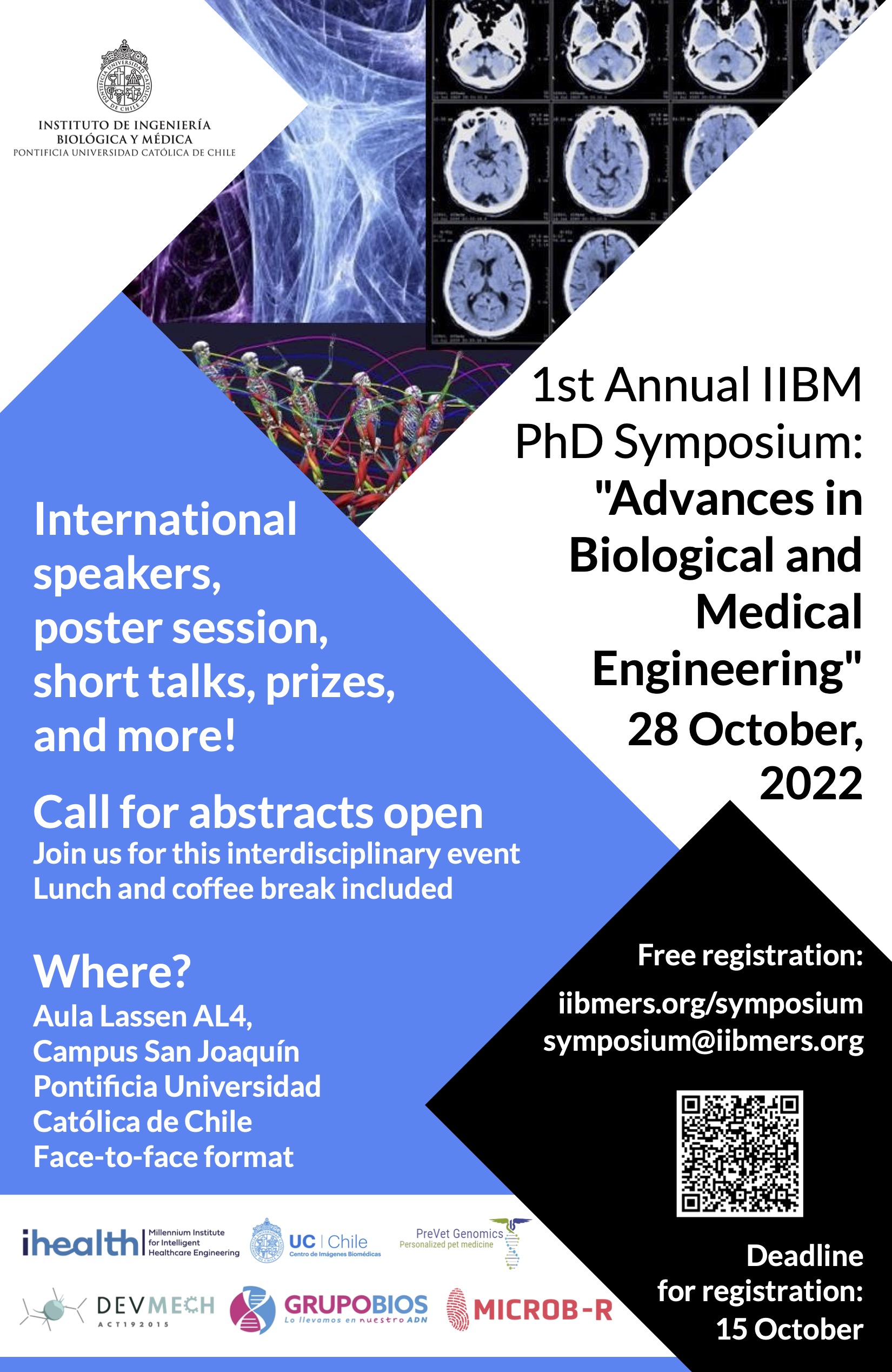
- Event date : 28 October 2022
- Cost : Free (includes certification for exhibitors and attendees)
- Abstract submission deadline : 17 October 2022 (12 PM UTC-3)
- Acceptance Notification : 20 October 2022
- More information : symposium@iibmers.org
- Starting time : 9:30 AM (UTC-3)
List of students’ contributions (grouped by topic):
> Protein engineering
Dimer dissociation is the rate-limiting step of KaiB fold switching - Ignacio Retamal (presentation & poster)
Cyanobacteria harbor the simplest biological clock, composed of three proteins (KaiA, KaiB, KaiC) that constitute a phosphorylation oscillator whose periodicity is regulated by the metamorphic protein KaiB. These proteins are characterized by adopting dissimilar yet thermodynamically stable structures, each with its own function. In the case of KaiB, a structural interconversion occurs between a tetramer of asymmetric dimers, or ground-state (gsKaiB), and a thioredoxin-like monomer, or fold-switch state (fsKaiB).
Although both structures and their functions are well described, how the structural interconversion occurs and what is its rate-limiting step are unknown. Here, we employ molecular dynamics simulations using structure-based models (SBM) with dual-basin Gaussian contact potentials to explore the fold-switch of KaiB. From these simulations, dimer dissociation is identified as the rate-limiting step of KaiB fold-switch, following a three-state refolding pathway with accumulation of a monomeric intermediate (gs2 ⇄ 2gs ⇄ 2fs). Lastly, the importance of oligomer stability for the fold-switch of KaiB is demonstrated by experimental analysis of two mutants with altered periodicity, for which a shift towards population of the dimer and monomer species and local structural features compatible with fsKaiB are observed by size exclusion chromatography and hydrogen-deuterium exchange mass spectrometry, respectively.
Altogether, these results suggest that biophysical analysis of the transition state for the structural interconversion between gsKaiB and fsKaiB can be exploited for rational design of the periodicity of the cyanobacterial circadian clock.
Biologic production of indigo dyes and derivatives - Nicolas Nuñez (presentation)
Indigo compounds have been widely used through the last centuries, from the Tyrian purple in the Mediterranean, even more expensive than gold, to the Asian cultures who used them as pigments and medicines. Nowadays, the most common use of these pigments is to dye jeans. Moreover, several studies have reported other applications as pharmacologically active compounds and as electronics components. The traditional way to produce them has been through extraction from natural sources (sea snails or plants), however they are currently synthesized through chemical procedures starting from aniline, a petroleum derivative. Here, we present a biocatalytic method to obtain the main indigoids, indigo and indirubin, and new substituted derivatives observing a change in their color properties. The research involves the use of two novel variants of the Baeyer-Villiger Monooxygenases enzyme, phenylacetone monooxygenase (PAMO), which has been mutated in specifics residues, allowing it to accept new substrates as substituted indols. The reaction was performed using recombinant E. coli overexpressing these new enzymes. Indigo and indirubin were directly obtained from whole-cell E. coli cultures. Substituted derivatives were obtained using free enzyme combined with a cofactor regenerating system, adding the indols as starting materials. These biocatalytic methods are safety and environmentally friendly, use minimal hazardous compounds, reducing the contamination to the environment. Moreover, these versatile pigments have a broad range of research and industrial applications.
“SCHEMA-RASPP: A versatile and convenient tool for the designed of optogenetic switches to regulate protein function” - Cyndi Tabilo (presentation)
The regulation of protein function has been studied using different approaches, including their engineering via fusion with other proteins that recognize a specific stimulus as an input to trigger a functional response as an output. A novel approach is the design of light-inducible switches, which present accurate spatial and temporal regulation of biological processes, including enzyme activity. Usually, structural modeling and molecular dynamics are required to determine the most appropriate insertion points, but this rational design approach is time-consuming and computationally expensive. Here, we used SCHEMA-RASPP, an algorithm that takes a multiple sequence alignment and one protein structure as input to design libraries of protein chimeras, as a fast and computationally inexpensive alternative for identifying appropriate insertion points for designing optogenetic switches. We first confirmed the validity of SCHEMA-RASPP to predict the switch insertion in readily available optogenetic switches of Src, Abl, and bRaf kinase, finding insertion points similar to those designed by molecular dynamics. Then, we used SCHEMA-RASPP on CK1δ, a human kinase of clinical relevance due to its malfunction being the causative of diseases and for which the switch insertion point is not a trivial decision, as a test case for further experimental work. Overall, our work establishes that SCHEMA-RASPP is a convenient strategy for engineering switches designed to control protein activity.
> Tissue engineering
Development of a Photosynthetic Hydrogel as Potential Wound Dressing for the Local Delivery of Oxygen and Bioactive Molecules - Rocío Corrales (poster)
The development of novel biomaterials to improve wound healing is a critical clinical challenge and an active field of research. As it is well described that oxygen plays a critical role in almost each step of the wound healing process, in this work, an oxygen producing photosynthetic wound dressing was generated, characterized, and further modified to additionally release other bioactive molecules. Here, alginate hydrogels were loaded with the photosynthetic microalgae Chlamydomonas reinhardtii, showing high integration as well as immediate oxygen release upon illumination. Moreover, the photosynthetic hydrogel showed high biocompatibility in vitro and in vivo, and the capacity to sustain the metabolic oxygen requirements of zebrafish larvae and skin explants. In addition, the photosynthetic dressings were evaluated in 20 healthy human volunteers following the ISO-10993-10-2010 showing no skin irritation, mechanical stability of the dressings, and survival of the photosynthetic microalgae. Finally, hydrogels were also loaded with genetically engineered microalgae to release human VEGF, or pre-loaded with antibiotics, showing sustained release of both bioactive molecules. Overall, this work shows that photosynthetic hydrogels represent a feasible approach for the local delivery of oxygen and other bioactive molecules to promote wound healing
Comprehensive characterization of mouse skin derived from regenerative versus reparative stages: a comparative analysis - Valentina Castillo (poster)
Tissue regeneration capacities vary significantly in the course of life. For instance, some organism like mammals can fully regenerate only until determined fetal stages, after that tissue repairs take place without reestablish normal tissue architecture and function. The high regenerative potential of embryos has been attributed to several factors, such as stem cells, immune system, inflammatory response, cell-response to specific growth factors, secretion of matrix-degrading proteases and the presence of particular extracellular matrix molecules upon damage. In order to contribute to better understand the changes that locally occurs in the transition between regenerative and reparative stages, in this work we carried out a comparative analysis between skin derived from mice at regenerative (E16.5) and reparative (6-8 weeks) stages. For this purpose, molecular, structural, histological, mechanical and functional analysis was performed. Here, we found that fetal skin composition differs not only in their molecular composition and distribution but also in their structure and functionality. Our results showed increased nuclei acid, collagen III and glycosaminoglycans content in regenerative versus reparative stages, while, differential lipid distribution, pore size and proteoglycans amount were observed too. Moreover, we also found that regenerative skin has significantly higher porosity, metabolic activity, fluid capacity and elasticity than reparative skin. This work provides a great knowledge about molecular, structural and functional hallmarks of skin derived from stages with regenerative abilities. Moreover, our results give insights for the development of new scaffolds with clinical potential able to mimic both structure and composition here described.
Development of self-assemble photosynthetic scaffolds by engineering microalgae to release human recombinant fibrinogen and thrombin. - Felipe Carvajal (poster)
Tissue engineering develops novel biomaterials that behaves and have functionality like natural tissues, with focus in improving the healing process after an injury. Such biomaterials should be porous 3D structures (scaffolds), be biocompatible, non-immunogenic, have an appropriate degradation rate, improve the tissue repair process, and host cells and cytokines from the regenerative process. Commercially available collagen scaffolds, used in tissue regeneration, are made of purified collagen from animal origin, which pose a risk of immune rejection on the host. An alternative could be the use of recombinant human collagen, but no appropriate biological platform has been used. Wounds imply the impairment of the vascular network, a problem not solved by any commercially available scaffold. The low oxygenation of the tissue impairs the regeneration process. In our laboratory we solved this issue, by delivering oxygen to the wound by using a photosynthetic collagen scaffold, including microalgae cells in the scaffold. This scaffold is fabricated over an inert commercially available collagen dermal matrix. To simplify the application and fabrication process, to improve the biocompatibility, and to provide a living biomaterial, a novel fibrin photosynthetic scaffold will be developed. Fibrin is a protein found in clots and serves as a natural scaffold in wounds. By producing two genetically modified strains of Chlamydomonas reinhardtii cells, that when combined produce fibrin, we will obtain a safe, functional self-assemble biomaterial, capable of self-regenerate and deliver oxygen on the application site, and could be used as platform for the delivery of other molecules to the wounds.
Towards photosynthetic explants as local oxygen delivery systems for tissue regeneration - Sergio González-Itier (poster)
Oxygen is necessary for tissue regeneration as it plays a key role in metabolism, immune response, and protein synthesis. Thus, the delivery of oxygen to wounds is an active field of research. Recently, in vitro, and in vivo studies have shown that photosynthetic biomaterials are a promising approach to deliver oxygen in a vascular independent manner. Due to its physical and chemical properties, plants have been used to improve tissue regeneration, but their capabilities to produce and deliver oxygen in situ have not yet been explored for wound healing. In this work, the influence of different environmental conditions in the oxygen releasing capacity of Marchantia polymorpha explants was characterized under human physiological conditions. Additionally, the biocompatibility of such explants as well as their capacity to fulfill the metabolic requirements of living vertebrate system was studied using a zebrafish model. Here we found that the explants were able to produce oxygen under human physiological conditions under light/dark cycles of 15 min. Moreover, co-incubation with zebrafish larvae demonstrates that M. polymorpha explants are not toxic, as viability of the larvae was not compromised. Finally, co-incubation assays also demonstrate that, under the appropriate illumination, the explants can exceed the oxygen demand of 96 hours post fecundation zebrafish larvae. Our proof-of-concept results suggest that photosynthetic explants could be used as biocompatible local oxygen delivery systems for wound healing and tissue regeneration. Nevertheless, further studies should be conducted in order to confirm the safety and efficacy of M. polymorpha to promote wound healing in vivo.
Photosynthetic oxygen production for tissue engineering: optimization and development of a new data analysis tool. - Pablo Rozas (poster)
Oxygen is critical for proper physiological functions including wound healing. Vascularization is compromised in full-thickness wounds and, thus, there is no proper oxygen supply from blood. Efforts have been made to treat patients with collagen scaffolds to promote wound healing. Nevertheless, oxygen availability is not optimal under these conditions. To cover this need, we have employed photosynthetic microorganisms to produce oxygen locally inside the scaffold. Here, we analyze light settings to optimize this oxygen production in terms of wavelength and intensity. Also, we developed a new oxygen levels data analysis platform to assess oxygen metabolism rapidly, reliably and user-independent. This tool is useful not only to study photosynthesis but also other metabolic parameters in biological systems such as mitochondrial respiration. Finally, this tool can be used by anyone interested in using oxygraph systems in their experimental procedures.
> Chemistry
Design and obtention of asymmetric diiminoacenaphthenes as potential ligands in Copper (I) complexes applicable to LEC devices and photocatalysis - Ignacio Erazo (poster)
In the permanent search for systems that allow the conversion of energy, either to emit light or favor chemical transformations, great advances have been made in the last decade. Moreover, various research groups around the world have studied the properties and characteristics of Ruthenium, Iridium and Copper complexes, both in light-emitting devices and redox photocatalysis. The heteroleptic complexes of Copper (I) type [Cu(N^N)(P^P)]+ have stood out for their attractive photoredox properties, which can be regulated through the structure of the ligands and their combination, affecting the absorption of visible light, the half-life of the excited state, stability in the reaction medium, electrochemical reversibility, synthetic facility, among others.
In this sense, ligands derived from symmetric diiminoacenaphthenes have been widely used in complexes, with several synthetic and photochemical applications, since it is considered a non-innocent redox ligand due to its strong electron-withdrawing properties. However, there are few reports of asymmetric derivatives and even fewer using heterocyclic substituents such as diazaborole derivatives, which have a strong electron-donor character. In this sense, this work shows the design, synthesis, and characterization of a new family of asymmetric diazaborolyl-diiminoacenaphthene ligands, and its potential use in the formation of Copper (I) complexes with applications in LEC devices and photocatalysis.
> Green chemistry
Bio-click chemistry: much more than play with Green Legos - Diego Fernando Rodríguez Sánchez (poster)
The fields of click chemistry and biocatalysis have rapidly grown over the last two decades. The development of robust and active biocatalysts and the widespread use of straightforward click reactions led to significant interactions between these two fields. Therefore, the name bio-click chemistry seems to be an accurate definition of chemoenzymatic reactions cooperating with click transformations. Bio-click chemistry can be understood as the approach towards molecules of high-value using a green and sustainable approach by exploiting the potential of biocatalytic enzyme activity combined with the reliable nature of click reactions. The present work summarizes the principal bio-click chemistry reactions reported over the last two decades, with a special emphasis on small molecules. Contributions to the field of bio-click chemistry are diverse, but the synthesis of chiral molecules with applications in medicinal chemistry and sustainable syntheses will be especially highlighted.
> Medical chemistry
What do we know about PFCs? - Francisco Castillo (presentation & poster)
PFCs or perfluorocarbons or perfluorochemicals are not widely known. Still, they are commonly associated with chlorofluorocarbons (toxic to the ozone), Teflon (known material for non-sticky kitchen utensils), or perfluorooctanoic acid (common manufacture byproduct of other commercial goods, which is a contaminant). PFCs have been studied in the biomedical area with a wide range of uses, but the most important one is their capability to transport low molecular weight gases, like oxygen. Starting with Clark’s Rat, in which a mammal survived many hours in an oxygenated perfluorochemical, multiple studies have been published, but very few of them studied the characteristics, properties, and behavior of the PFCs and their interaction with oxygen. It is known that PFCs are lipophobic and hydrophobic at the same time and that the bonds between carbon and fluor affect the structure when compared with their hydrocarbon homolog, but… What do we know about PFCs? In this study, we took a simple perfluorocarbon and studied its structure, energetic stability, electronic parameters, and its interaction with oxygen, using theoretical-computational chemistry, in order to understand its behavior and capability to capture oxygen, in the search for the most suitable PFC to use in bioapplications as oxygenating systems for oxygen therapy or organ transplantation.
Molecular modeling study on allosteric regulation of Cannabinoid Receptor Type 1 by Cannabidiol-induced lipid bilayer membrane perturbations - Hery Chung (poster)
Within the endocannabinoid system (ECS) the transmembrane cannabinoid receptor type 1 (CB1) regulates a variety of physiological and pathological conditions constituting a promising therapeutic target. Various mechanisms are involved in CB1 signaling and together with ligand binding responses, the lipid composition of the surrounding bilayer can represent an additional source of protein regulation. In the light of growing evidence that relates the phytocannabinoid cannabidiol (CBD) and endogenous cholesterol (CHL) through a possible common binding site and associated metabolic pathways, CHL may be regarded as an integral part of CBD’s action. In this way, changes in membrane sterol content could be intimately related to CBD’s effects and together modulate receptor structure. This project aims to explore how membrane CHL can indirectly modulate receptor function and take part in the described allosteric effects of CBD. We propose that together with direct allosteric binding, changes in membrane CHL in the presence of CBD, can also favor an inactive-like state receptor and contribute to the reported negative modulatory effects. A molecular simulation approach combining protein modeling, molecular docking and dynamics simulations with all-atom (AA) and coarse-grained (CG) methodologies will be used to generate structural and energetic data of representative receptor systems. Exploring the membrane contribution to CBD’s effects can help to gain better understanding of its mechanisms of action and provide deeper insight into transmembrane protein modulation. Given the therapeutic potential of CB1 and the positive clinical effects shown by CBD, research on its underlying mechanism will be useful to guide medicinal chemistry efforts.
> Drug delivery
DESIGN AND OPTIMIZATION OF MULTIFUNCTIONAL NANOSYSTEMS FOR NEEDLE-FREE VACCINATION. - Rayen Valdivia (presentation & poster)
Vaccination is one of the most significant milestones in public health worldwide. According to data from the World Health Organization, there are currently vaccines for more than 20 life-threatening diseases, preventing around 3 million deaths yearly. However, manufacturing costs, logistics, supplies, and risks associated with the traditional vaccine administration route (parenteral), contribute to the vaccination coverage gap. Scientists have successfully developed safer but less immunogenic antigens (necessitating an adjuvant). However, more efforts are still required to develop adjuvants and increase their bioavailability. Nanomedicine allows the development of new systems capable of crossing biological barriers, turning them into novel platforms for antigen delivery by needle-free routes. This work aims to develop colloidal systems with virus-like sizes capable of jointly encapsulating a model antigen and an active molecule with adjuvant properties. This multifunctional approach allows having a formulation able to transport the antigen to the immune system and activate it due to its adjuvant activity. Different formulations have been optimized through mathematical modeling to modulate their physicochemical properties to respond to the challenge of crossing the skin barrier. The new nanosystems have sizes ranging between 50 and 700 nanometers, a monomodal population of particles and surface charges that ensure the stability of the systems. Optimal formulations are currently undergoing stability and antigen plus adjuvant association studies simultaneously. Subsequent ex vivo studies in pig skin will allow us to evaluate this platform as needle-free formulation and its potential contribution to the global vaccination.
Natural Killer cell-derived exosome mimetics as an alternative for encapsulation and drug delivery in lung cancer cells. - Javiera Carrasco (poster)
Lung cancer (LC) has the highest mortality rate worldwide. The pathogenesis is multifactorial and targeted therapies are currently recommended treatment; however, patients who don ́t qualify for this therapy must resort to classic treatments (e.g. Chemotherapy), although it has limitations (e.g. side effects, chemo-resistance, etc.) Exosome mimetics (EM), artificially generated vesicles with exosome properties, have been proposed as a potential tool to lower barriers for clinical translation. We propose a formulation of a chemotherapeutic encapsulated in EM generated from natural killer (NK) cells (EM-NK-C) establishing their cytotoxic effects on LC. Methodology: EM-NK-C were generated by cell extrusion. Morphology was analyzed using atomic force microscopy. Quantification was performed by nanoparticle tracking analysis. Determination of NK and exosome markers were performed by western blot. The cytotoxic effect was determined by MTT in NCI-H1299 and NCI-H1975 cell lines. Results: The mean size of NK exosomes (EXO-NK), EM and EM-NK-C were within exosome range (<200nm). The presence of all the markers analyzed in the EM and cell lysate was determined, in contrast to the EXO-NK, which didn ́t present calnexin and GAPDH. For both cell lines a tendency of dose-response effect is observed when exposed to ratios of 1:1, 1:10 and 1:100 (vesicles:cells). Conclusion: We established a methodology to generate EM and EM-NK-C. Although, so far, we haven ́t been able to establish a cytotoxic effect, further experiments must be carried out to complement these results. Acknowledgements: We would like to thank Dr. Margarita Montoya (Universidad de Santiago de Chile) for kindly donating NK cell line.
Exploring the applications of a new copolymer in nanoparticle synthesis - Barbara Astudillo (poster)
Diverse research has reported the application of polymers in tissue regeneration, especially related to the regeneration of damaged skin. In this field, our group describes a novel method to produce a new biopolymer (patent pending). This biopolymer was designed to generate a biomaterial that could be used not just to enhance the regeneration of the tissue but also be apply in the development of different systems such as nanosystems. In this work, we present an in-progress application of the copolymer where we explore its application in the generation of nanoparticles with diverse core molecules. Our result shows that we can generate nanoparticles with different nucleus core molecules. Additionally, the obtained results showed that the hydrophobic nature of the core molecule is a critical factor in the formation of the nanoparticle from this new copolymer.
Innovative techniques for the Nanoworld: evolution in Lipid Nanoparticles formulation. - Karla López (poster)
The dissolution and absorption of lipid active pharmaceutical ingredients (API) into the human body represents a challenge for its administration due to their low solubility in water. In order to solve this problem, it is necessary to improve the solubility and the delivery to therapeutic target. Considering this challenge, nanomedicine represents an alternative focus on the formulation of different type of nanocarriers allowing the administration of complex API, and preventing the degradation of the molecules, through their encapsulation, offering better biodistribution. Nevertheless, some nanocarriers as metallic ones have shown to be associated to different cytotoxic effects. That is the reason why a new generation of lipid nanovehicles have emerged, known as Solid Lipid Nanoparticles (SLN) and Nanostructured Lipid Carriers (NLC). Our research is based on the designing of novel SLN or NLC with biocompatible raw materials, ideal to encapsulate lipid molecules, and avoiding the use of organic solvents. To reach the production of SLN and NLC, we have been applying different methods with moderate amounts of external energy. Additionally, we are testing the use of green chemistry techniques such as: Microwave-Assisted Synthesis (MAS), that have currently been adapted for the manufacture of said nanocarriers. This method involves fewer and quicker steps than traditional ones, allowing the nucleation and particles formation. Our formulations have shown smaller nanometric sizes, lower polydispersity index and higher API solubility than those obtained by conventional methods. This currently in progress research will show the additional advantages of these novel nanocarriers in the new era of nano-formulation.
Zr-based MOF as drug delivery system for drugs - Javier Salazar (poster)
In general, low bioavailability, high side effects, and rapid clearance from the body are typical problems related to the therapeutic application of different agents. Delivery systems search to solve these problems through a noninvasive approach. Metal-organic frameworks (MOFs) are crystalline microporous characterized by presenting a net-like structure composed of a metal atom linked through organic ligands. MOFs have small sizes, high surface areas, and increased drug-loading capacity. However, the application of MOF in the liberation of the therapeutic agent presents some challenges due to the heavy metals used in their structures and difficulties in controlling drug loading and release. In this work in progress, we study the capacity of a Zr-based MOF to load and release a well-described drug such as diclofenac. We used a structural approach to understand the effect of the drug on the structure of the MOF. We tried to elucidate how we can improve the system’s stability, loading, and release by enhancing the system’s design.
> Biomedical imaging
Banding artifact correction in DESPOT2 using a theoretical dictionary approach - Ronal Coronado (presentation)
DESPOT2 is mri technique to quantify T2 maps. But, DESPOT T2 is based on balanced steady-state free precession (bSSFP) acquisition which is sensitive to banding artifacts. In our work, we propose a theoretical banding correction using a dictionary of magnetic fingerprints. This dictionary takes into account the Bloch simulations for high combinations of T1, T2, and off-resonance. The results in simulations and phantom acquisitions show a considerable improvement in the quality of T2 maps in comparison to classical DESPOT reconstruction. This approach could allow to apply DESPOT2 in any region of the body, independently of the off-resonance strength.
Intensity-based Deep Learning for SPION concentration estimation in MR imaging - Alberto Di Biase (presentation & poster)
We present a new method for estimating the concentration of Superparamagnetic Iron Oxide Nanoparticles (SPION) in magnetic resonance imaging (MRI). SPION have a wide range of biomedical applications and use as a contrast agent in MRI. When used in MRI, SPION produce off-resonance effects in MRI, as distortions of the phase and magnitude, which can be used to estimate their concentration. At high concentrations, these artifacts can be severe and cause signal loss, making the phase information unusable. By focusing only on the magnitude or intensity distortions in the surrounding tissue and using a neural network, we can find the concentration of SPIO. In contrast to previous methods which rely on the phase or take multiple images, ours uses only two magnitude images, reducing imaging time. The network input is the difference of the 3D intensity volumes and the output is a map with the SPION concentrations. Instead of using a 3D architecture, we use a 2D U-net with filters as the third dimension. It was trained with simulated data. We tested our method with simulated images (NMSE: 0.3838), with numerically inserted SPION in actual breast images (NMSE: 0.7355), and with phantom data obtained from a 7T scanner.
Quantitative Susceptibility Mapping MRI in deep brain nuclei in first episode psychosis - Marita García Saborit (presentation & poster)
Background: Psychosis is related to neurochemical changes in deep-brain nuclei, particularly suggesting dopamine dysfunctions. We used an MRI-based technique called quantitative susceptibility mapping (QSM) to study these regions in psychosis. QSM quantifies magnetic susceptibility in the brain, which is associated with iron concentrations. Since iron is a co-factor in dopamine pathways and co-localizes with inhibitory neurons, differences in QSM could reflect changes in these processes. Methods: We scanned 83 patients with first episode psychosis and 64 healthy subjects. We reassessed 22 patients after three months. Mean susceptibility was measured in six deep-brain nuclei. Using linear regressions, we analyzed relationships between QSM and case-control differences, gender, volume, treatment duration, dose, laterality and psychotic symptoms. Results: Patients showed a significant susceptibility reduction in putamen, thalamus, and globus pallidus externa (GPe). Patients also showed a significant R2* reduction in GPe. After 3 months of antipsychotic treatment, patients showed significant changes in symptoms and susceptibility in globus pallidus interna and thalamus. We also found that positive PANSS score is a predictor variable of susceptibility changes in Substantia Nigra. Gender, laterality, dose, treatment duration, and volume were not predictor variables of QSM. Conclusions: Reduction in QSM and R2* suggests a decreased iron concentration in GPe of patients. Susceptibility reduction in putamen and thalamus cannot be associated with iron changes. Since changes observed in putamen and GPe were not associated with symptoms, dose, and treatment duration, we hypothesize that susceptibility may be a trait-marker rather than a state-marker, but this must be verified with long-term studies.
Liver MRI-PDFF estimation using a two-stages Variable Echo-Times Neural Network (VET-Net) - Juan Pablo Meneses (presentation & poster)
MRI Proton Density Fat Fraction (PDFF) is a relevant and extensively validated biomarker associated to Non-Alcoholic Fatty Liver Disease (NAFLD). Recent literature has proposed several Deep Learning (DL)-based approaches to estimate PDFF from multi-echo gradient echo images. The main advantage of DL-based techniques over reference methods is the acceleration of the overall procedure (shorter MR scans + faster post-processing) without a significant diminishing of performance. However, the robustness and reproducibility of DL-based methods for PDFF quantification had not been extensively assessed. In this work, we propose a novel two-stages and echo-times (TE)-aware convolutional neural network, denoted as Variable Echo-Times Neural Network (VET-Net) that is capable of estimating PDFF maps from multi-slice abdomen MR images. The two-stages procedure consists of a first computation of R2* and off-resonance field maps, which are confounding variables associated to the fat quantification process, to posteriorly estimate water and fat concentrations using and ordinary least squares method. Additionally, a vector with TEs is incorporated as a second input to VET-Net by using adaptive instance normalization (AdaIN) layers that modify the extracted features according to the received input vector. VET-Net performance was assessed in terms of bias, considering a multi-site multi-vendor phantom dataset, and precision, using subjects acquired several times using different TEs. Results showed a significantly improved performance compared to already proposed DL-based techniques, which placed VET-Net as a highly robust and reproducible technique to estimate PDFF.
A desktop low-field MRI: Current progress and future steps - Guido Muñoz (presentation & poster)
A low-field model for a magnetic resonance device is presented. It’s designed to illustrate, at a “nuts a bolts” level, how magnetic resonance works, it’s main parts, and the role which hardware plays into it. which is often overlooked in favour of imaging techniques by software tinkering. Currently it has a B0 field of 0.12 T, B1 pulses of approximately 100 uT and a field of view of 2.1 mm. It is currently able to produce an FID of low intensity. Current design choices are presented, alongside possibilities for future development and possible future applications.
Breast tumor volume quantification from Digital Breast Tomosynthesis (DBT) using a Deep Learning approach - Cristina Alfaro (presentation & poster)
In 2020, breast cancer has become the leading cause of cancer in women worldwide. Thus, early detection of suspicious lesions is essential to improve prognosis and avoid radical mastectomies. Digital 2-D mammography (DM) has been the gold standard to assess early breast lesions but has important limitations due to tissue overlapping. This difficult the detection of suspicious lesions especially in patients with dense breast tissue. Recently, Digital Breast Tomosynthesis (DBT), a pseudo-3D breast imaging technique has shown the potential to provide a volumetric set of 60 to 70 thin (1 mm) slices acquired through the rotation of an x-ray tube in an arc of narrow angle above the compressed breast tissue, reducing the effect of tissue overlapping and improving the capacity to visually distinguish between the normal radiographically dense breast tissue and malignant lesions. Despite the evident benefits of using DBT, several studies have shown contradicting results for the tumor size assessment as compared to histopathologic measurements. Both procedures are traditionally performed manually or through semiautomatic techniques. In other hand, Deep Convolutional Neural Networks (CNNs) models have the advantage of being operator-independent and extract information from the DBT images. This research aims to use a Deep Convolutional Neural Network-based approach to accurately quantify the volume of irregular-shaped breast tumors from DBT images. This will contribute to improve the assessment of size, morphology, and spatial location relative to surrounding healthy breast tissues, especially in clinical settings where imaging modalities with full 3D capabilities such as MRI are not available.
Focal volume microscopy for high-speed three-dimensional optical neurophysiology - Maximiliano Valdes (presentation & poster)
Understanding mechanisms of how the brain works –or stops working– will allow more precise diagnosis and treatment of human neurological and psychiatric diseases. Zebrafish larvae swim against flowing water to maintain their location in familiar territory, for which they remember their visual environment over many seconds. Their memory and its localization in the brain have been studied by imaging slow persistent neuronal Calcium transients upon patterned visual stimulation. The time resolution of current cell-resolved volumetric neuronal activity recordings does not allow resolving whether the persistent activation is sustained by an intrinsically slow activation or by a fast-recurrent network, even though faster fluorescent reporters of synaptic release and of membrane voltage are available.
To address this question, we propose to develop a new class of microscope and algorithms to enable fast, localized optical neurophysiology of live behaving zebrafish. We aim to record neuronal activity at 50 Hz bandwidth from a 700x500x110 μm3 volume with cellular resolution. Unlike existing cell-resolved light-sheet microscopes that only resolve up to 5 Hz, or existing fast volumetric microscopes that compromise cell resolution, our original approach to volumetric recording is multi-focal virtual image formation with a single camera. Each frame captures an array of virtual images focused at different depths of the sample, generated by reflections inside an optical cavity. The virtual array imposes different path lengths onto sections of the imaging aperture, thereby allowing simultaneous focusing at multiple depths on a volumetric sample while maintaining a diffraction-limited resolution.
Construction of a high spatio-temporal resolution volumetric microscope using multiplane imaging. - Maximiliano Mariné (presentation)
In recent years, the zebrafish has been used as a model to study memory and visual information integration circuits. Current methods of volumetric imaging of neuronal activity lack sufficient temporal resolution to distinguish whether persistent activations of memory-associated areas are due to slow activations or to recurrent integrating circuits with fast activations. To solve this problem, we propose a novel kind of volumetric microscope that captures multiplane images in a single shot. In this work we built and characterized the first prototype of this microscope, designed to record neural activity in a volume of 700x500x110 μm3 at a temporal resolution of 50 Hz to allow rapid probing of the dynamics of the whole zebrafish brain. We built a custom microscope using a Nikon 10x objective and achromatic secondary lenses resulting in a standard image with 5x magnification, and captured multiplane images with a digital camera. Design parameters were calculated using MATLAB simulations and theoretical analysis. An USAF 1951 test target was translated axially to evaluate its defocusing in each image. We characterized multiplane imaging configurations with 3 and 4 planes, with an approximate separation of 15 µm between planes, and FOV of 690 and 500 µm respectively. These results demonstrate the feasibility of the prototype to capture single-shot multiplane images.
Progress on all-optical neurophysiology to study neuronal activity in brain tissue at IIBM - Ruth Vallejos (poster)
Widefield all-optical neurophysiology uses optical stimulation and recording of neural activity to characterize neuronal function over large areas of brain tissue. For this purpose, a fluorescence microscope with large FOV (4.6 mm) and cellular resolution is used to stimulate optogenetic actuators and measure activity reporters simultaneously in different color channels. To stimulate and record activity from thousands of neurons with one photon (1P), a blue-excited channelrhodopsin (ChR2) is paired with a red-shifted calcium indicator (H2B-jRGECO1a). To record activity from individual neurons over large areas of brain tissue, a digital micromirror device (DMD) is used to project patterns and illuminate neighboring sample locations with orthogonal functions of time based on Hadamard codes, uncorrelated background is rejected computationally. These tools can be used for wide-area interrogation of neuronal excitability and functional connectivity in acute brain slices. Here we report progress on the second-generation implementation of this microscope, based at the IIBM in the Faculty of Biology. This new microscope version will include a zoom function to tailor the projection and imaging FOVs to different samples, and design updates to facilitate modular implementation with simpler and lower cost components. We currently have started configuration and assembly of the equipment. We plan to fully test the operation of the system by the end of this year. This tool will make it possible to study the interaction of neurons in brain tissue to better understand their involvement in mental health disorders, and will open the opportunity to use all-optical neurophysiology in future collaborative projects.
Scanning angle interference microscopy to analyze protein nanoscale position and orientation - Nicolas Miller (poster)
For many years optical microscopy has served humankind to study the microscopic world, nevertheless, it has a limit in what we can observe, that is affected by the famous Abbe limit of diffraction (to approximately 200 nm in xy-dimensions and 500 nm along the optical axis.). This limit poses a problem when trying to image subcellular structures. To overcome this obstacle, in the past decade, various methods of superresolution microscopy have been developed that completely shattered the diffraction limit of image resolution. Here we are presenting a method based on interference contrast, which is known as scanning angle interference microscopy (SAIM). This technique can be easily optimized on a TIRF microscope and allows for localization with nanometer precision along the z-axis. This allows to extract information about the position and orientation of proteins in the z axis, therefore, it is a convenient and very useful tool to image the vertical positions of cellular structures, with a minimal sophistication and to image protein dynamics in living cells.
> Mechanobiology
A flexible computational framework of cell aggregation identifies minimal mechanical interactions for onset of multicellularity in early annual killifish embryogenesis - Ignacio Montenegro (presentation & poster)
Deciphering the biological and physical requirements for the outset of multicellularity is limited to few experimental models. The early embryonic development of annual killifish represents an almost unique opportunity to investigate de novo cellular aggregation in a vertebrate model. As an adaptation to seasonal drought, annual killifish employs a unique developmental pattern in which embryogenesis occurs only after undifferentiated embryonic cells have completed epiboly and dispersed in low density on the egg surface. Therefore, the first stage of embryogenesis requires the congregation of embryonic cells at one pole of the egg to form a single aggregate that later gives rise to the embryo proper. This unique process presents an opportunity to dissect the self-organizing principles involved in early organization of embryonic stem cells. Indeed, the physical and biological processes required to form the aggregate of embryonic cells are currently unknown. Here, we developed an in silico, agent-based biophysical model to test how cell-specific and environmental properties could determine the aggregation dynamics of early Killifish embryogenesis. In a first approach (cell autonomous system), we considered how intrinsic biophysical properties of the cells such as motility, polarity, density, and the interplay between cell adhesion and contact inhibition of locomotion drive cell aggregation into self-organized clusters. Second, we included guidance to cell migration through a taxis mechanism to resemble the activity of an organizing center inbuilt in the embryo environment. Our numerical simulations showed that random migration combined with low cell-cell adhesion is sufficient to maintain cells in dispersion and that aggregation can indeed arise spontaneously under a limited set of conditions following characteristic power laws, but, without environmental guidance, the dynamics and resulting structures do not recapitulate in vivo observations. Thus, an environmental guidance cue seems to be required for correct execution of early aggregation in early killifish development. However, the nature of this cue (e.g., chemical or mechanical) can only be determined experimentally. Our model provides a predictive tool that could be used to better characterize the process and, importantly, to design informed experimental strategies.
Microfabricación de dentina biomimética como sustrato para explorar adhesión de biopelículas in-vitro - José Morales (poster)
La dentina, el tejido más abundante del diente humano, se ubica en una capa inferior al esmalte y cubre el tejido pulpar. En su estructura presenta túbulos que rondan los 4 um de diámetro de separación variable, que están directamente asociados a su homeostasis y enfermedad. Por ello, es importante buscar nuevos modelos in-vitro de estudio de estas condiciones dentro del laboratorio. La microfabricación es el proceso utilizado para fabricar objetos con dimensiones nanométricas para el estudio de sistemas biológicos y recrear superficies, sin embargo, en la literatura existen escasos reportes del uso de estas técnicas para el diseño de dentina biomimética. El objetivo de este estudio fue diseñar y generar una dentina biomimética in-vitro a través del método de fabricación con resina. Se generó un diseño de microestructuras mediante el software Klayout, con el cuál se confeccionó una superficie con micropilares de resina por medio de la técnica de fotolitografía. Las medidas de los pilares tuvieron un diámetro de 5 um con separación de 6 um, 8 um y 10 um entre ellos. Posteriormente, con el patrón obtenido del wafer, se generó un patrón negativo de polidimetilsiloxano (PDMS), donde se le realizó una cubierta de colágeno tipo I. Las superficies fueron visualizadas mediante microscopía óptica y electrónica de barrido. Finalmente, se logró confeccionar un wafer con las dimensiones requeridas, con el cuál se generaron superficies de PDMS y colágeno tipo I, observadas con microscopía óptica y electrónica. Se espera que estas superficies sirvan de sustrato para futuros experimentos.
Vinculin is the pivot of a novel ternary complex mediating force transduction at adherens junctions - Nicole Morales (presentation & poster)
The multicellular tissue organization is mediated by a mechanical force transmission process controlled by a high-regulated adhesive complex called adherens junction (AJ). AJ proteins are compartmentalized into three main layers for signaling, force transduction and actin regulation. In the mechanotransduction layer we can find proteins such as catenins and vinculin, that sense and bear mechanical forces transmitted between intercellular contacts and the actin cytoskeleton. However, the molecular organization and arrangement of these components still remains elusive. Using super-resolution microscopy, biochemistry assays, and in silico simulation, we report that vinculin, once activated, could form a direct structural connection with β-catenin, which can even bypass α-catenin, probably the most known mechanotransducer in AJs. The results of experimental assays indicate that this vinculin/β-catenin interaction can support mechanical tension and conduit the connection of the cadherin-catenin layer to the actin cytoskeleton. Likewise, the molecular dynamics simulation show that, in the presence of α-catenin, vinculin/β-catenin interaction could be the means for the formation of a new ternary complex centered around vinculin, with simultaneous bindings to α-catenin and β-catenin to reinforce the AJ. These findings suggest a multi-step model for the force transduction at AJs whereby α-catenin may serve as the initial catalytic activator for vinculin, followed by the insertion of β-catenin to form a ternary complex contributing to the generation of the graded response needed in tissue homeostasis.
Development and Prototyping of a Magnetic Tweezer to Investigate Cell Mechanics - Matias Eduardo Moreno Fuenzalida (presentation & poster)
Investigation of mechanical features of cells is important to understand fundamental aspects of biological processes, physiological functions and onset of pathological conditions. In this context, optical and magnetic tweezers proved a powerful tool to mechanically stimulate cells via precise control of microspheres interacting with the cell. In this interdisciplinary project, we have design, fabricate and text a cost-effective prototype of magnetic tweezers that can provide fast and controllable actuation of a magnetic field. Our project consists of several steps and various geometries, materials, and feedback control systems are being designed and tested in order to produce accurate manipulation of the magnetic field. As a proof of concept, we have successfully tested a first prototype using magnetotactic bacteria that, under specific conditions, are capable of moving along the magnetic field. Currently the major challenge of our project is the disruption of global supply chain and still some materials are kept on hold. In this poster, we will present our mechanical and electronic designs, preliminary results and discuss future steps.
Wound Model Assay to study collective cell migration of epithelial cells. - Patricio Canales (presentation & poster)
Collective cell migration is of fundamental importance for embryonic development, tissue regeneration, wound healing and cancer metastasis. Regulation of large-scale collective migration depends on the interaction between cellular forces and regulated adhesion to neighboring cells and the substrate. However, the relationship between these mechanisms involved during collective dynamics, is still unknown. Here we have performed high resolution analysis of adhesion proteins and of actin architecture in an epithelial tissue (MDCK cells) using a Wound Model Assay (WMA) at 0, 4 and 10 hours from the onset of cell migration. In these experiments, we observed that there is an accumulation of Actin and E-cadherin at the leading edge at 10 hours as compared to 0 and 4 hours. This is related to the appearance of stress fibers perpendicular to the cell membrane in lamellipodia, and E-cadherin at the adherens junctions as well as in cytosolic vesicles. These preliminary results suggest that Actin and E-cadherin play an important role in the transmission of forces from leading edge to rear, contributing with traction force necessary for crawling through focal adhesions, and maintenance of tissue integrity through adherens junctions, respectively.
Analysis of possible mechanical features driving early embryonic development of A. nigripinnis using engineered biomimetic environments. - Sebastian Vásquez (presentation & poster)
Early annual killifish (A. nigripinnis) development is a unique model in vertebrates to study the mechanics of directed cell migration and de novo cell aggregation. Indeed, proper initiation of killifish development requires that embryonic stem cells (blastomeres), that are initially dispersed throughout the surface of the embryo moving by random walk in a confined space, begin a directed motion through an unknown guidance mechanism of biochemical or mechanical nature. Thereafter, cells coalesce into a single cluster that will eventually enter gastrulation to proceed with tissue differentiation and organogenesis. Due to the complexity of the system, investigation of cell migration directly in the native in vivo condition of the embryo presents several challenges including optical inaccessibility and difficulty in controlling environmental conditions. Hence, we developed an in vitro approach based on biomimetic engineered environments to investigate blastomere cell migration under controlled conditions using high resolution microscopy. Here, I will show our results demonstrating that (i) cellular activity varies in blastomeres extracted from embryos at different stages and that (ii) cells have preferential adhesion and enhanced activity when interacting with cell-cell junction adhesion molecules (E-cadherin) molecules. Finally, I will discuss our preliminary results that show how mechanical stimulation through confinement and compression could provide guidance for blastomere directed migration.
Bacteria swimming in complex fluids - Hector Urra (poster)
In nature, many bacteria are motile in environments of very different rheological properties such as Newtonian , viscoelastic or complex yield stress fluids. The ability for microorganisms to swim or eventually to be blocked, is an important practical issue sometimes related to serious health threat problems.For example, in medicine, bacteria can penetrate mucus protective barriers along the intestinal track and create inflammatory conditions or in the field, microbes can contaminate groundwater reservoirs out of septic tanks. To understand from a fundamental point of view, the problem of bacterial motility in complex fluids, I have studied the swimming and the exploration properties of a motile E. coli bacterium inside different model non-Newtonian fluids, such as carbomers or mucine gels. To characterize the motility in such media, I used a 3D Lagrangian tracking technique which allowed to obtain the 3D swimming trajectories of the bacteria. I observed and characterized how the swimming behavior is modified as a function of the mechanical interactions between the fluid and the swimmer. I also characterized the influence of local mechanical heterogeneities which create transient trapping and reorientation. Out of these measurements I defined the concept of “medium assisted tumbling” which progressively controls the large scale exploration features in a gel forming fluid. These results are promising and at the same time, of great help to understand, the mechanisms of penetration of bacteria inside the intestinal mucus. Along those lines, I obtained preliminary results in native porcine mucus extracts, in collaboration with a team of the Sorbonne University medical school.
The effect of Streptococcus sanguinis extracellular vesicles (EVs) on Streptococcus mutans - Camila Solar (poster)
Introduction: Dental caries are a relevant public health problem that is characterized by lesions on the dental surface that is associated with the presence of Streptococcus mutans (Sm) on the dental surface. Streptococcus sanguinis (Ss) has been associated with caries-free individuals and has shown an inhibitory effect to Sm. Extracellular vesicles (EVs) are lipid-bound structures that are secreted by cells in order to communicate with others. The purpose of this study is to evaluate if Ss extracellular vesicles can inhibit Sm growth.
Methods: We isolated EVs from Ss, Sm and a co-culture of Ss and Sm (Ss/Sm) through ultracentrifugation and characterized their size and shape by Nanotracking analysis (NTA) and transmission electron microscopy (TEM), respectively. We evaluated the effect of Ss, Sm, Ss/Sm and Ss+Sm EVs in Ss and Sm growth by growth curves.
Results: EVs samples were successfully isolated with an average size 161.9 ± 29.1 nm and 133.1 ± 39.8 nm for Ss and Sm. All the isolated EVs promoted Ss and Sm growth in a dose-dependent manner.
Conclusion: Our current protocol produces EVs that have a positive effect on Sm and SS growth. Changes in the culture medium may challenge Ss to produce EVs with inhibitory effect towards Sm.
Effect of changes in the physical properties of the extracellular matrix on autophagy - Beatrice Fantuzzi (poster)
Autophagy is a form of cellular housekeeping for degradation of proteins, organelles and other cellular materials Several environmental stresses activate autophagy, among those hypoxia, DNA damage, and metabolic challenges such as starvation. In addition to these chemical stresses, there is a requirement for cells to cope with their mechanical microenvironment. Cells accomplish this task through mechanosensitive protein complexes which interface the cells with their mechano-environment and with the cooperation of the cytoskeleton. Still the role of physical forces in autophagy regulation and their potential implications in both physiological as well as pathological conditions. With this investigation we want to understand the effect of changes in rigidity of the extracellular matrix on the autophagic flux. By using epithelial cells (MDCK) seeded on engineered biomimetic substrates with different rigidities, we are evaluating the appearance of LC3 and p62 carrying vesicles, and the organization of the actin cytoskeleton in response to serum starvation. This will allow us to analyze the differential activation of autophagy in response to mechanical stress.
> Biofilm mechanobiology
Microfabricación de dentina biomimética como sustrato para explorar adhesión de biopelículas in-vitro - José Morales (poster)
La dentina, el tejido más abundante del diente humano, se ubica en una capa inferior al esmalte y cubre el tejido pulpar. En su estructura presenta túbulos que rondan los 4 um de diámetro de separación variable, que están directamente asociados a su homeostasis y enfermedad. Por ello, es importante buscar nuevos modelos in-vitro de estudio de estas condiciones dentro del laboratorio. La microfabricación es el proceso utilizado para fabricar objetos con dimensiones nanométricas para el estudio de sistemas biológicos y recrear superficies, sin embargo, en la literatura existen escasos reportes del uso de estas técnicas para el diseño de dentina biomimética. El objetivo de este estudio fue diseñar y generar una dentina biomimética in-vitro a través del método de fabricación con resina. Se generó un diseño de microestructuras mediante el software Klayout, con el cuál se confeccionó una superficie con micropilares de resina por medio de la técnica de fotolitografía. Las medidas de los pilares tuvieron un diámetro de 5 um con separación de 6 um, 8 um y 10 um entre ellos. Posteriormente, con el patrón obtenido del wafer, se generó un patrón negativo de polidimetilsiloxano (PDMS), donde se le realizó una cubierta de colágeno tipo I. Las superficies fueron visualizadas mediante microscopía óptica y electrónica de barrido. Finalmente, se logró confeccionar un wafer con las dimensiones requeridas, con el cuál se generaron superficies de PDMS y colágeno tipo I, observadas con microscopía óptica y electrónica. Se espera que estas superficies sirvan de sustrato para futuros experimentos.
> Microbiology
Carbohydrate Metabolism in Bioleaching Acidophile Bacteria - Víctor Alegría-Mera (presentation)
Carbohydrates play a pivotal role in living creatures. In bacterial biofilms, carbohydrate metabolism is an essential process that ensures not only energetic purposes but also the production of extracellular polymeric substances (EPS), a variety of building blocks needed for the success and development of this sessile lifestyle. Although carbohydrate metabolism is a large and very depth subject that has been covered because of the importance of bacterial biofilms in health issues, little is known about this process in bioleaching acidophile bacteria and even less in the biofilm formation of these organisms. Bioleaching bacteria’s environment has highly acid pH values; the pH value of those places such as acid mine drainages (AMD) make it seemingly impossible to trigger a heterotrophic metabolism because of the lack of carbohydrates in there. To ensure energetic metabolism in these highly acidic environments, these bacteria fix and uptake the environmental CO2 present using the Calvin cycle and reverse TCA respectively. These microorganisms also use an incomplete TCA cycle, mainly for the biosynthesis of precursor molecules of interest, such as sugars, amino acids, and lipids, but in terms of the presence of carbohydrate transport systems or modification systems such as Glycosyltransferases (GTs), components required for the success of bacterial carbohydrate metabolism remains unknown. By in silico approaches, we aim to address the presence of carbohydrate transport systems and GTs as well as a set of possible carbohydrates that could be uptake by these bacteria during mineral attachment.
Caracterización de cepas multirresistentes de Escherichia coli aisladas desde verduras, aguas superficiales y personas de la ciudad de Molina, Chile: Un modelo de “Una salud” - Constanza Díaz (presentation & poster)
Las bacterias multirresistentes (MDR) a antibióticos están presentes en diversos ambientes y cada vez son más las vías de transmisión hacia las personas. El objetivo del estudio es caracterizar fenotípica y genotípicamente cepas de E. coli MDR aisladas desde diferentes matrices provenientes de la ciudad de Molina en Chile. Las matrices estudiadas fueron individuos sanos (n=143), vegetales (n=92) y agua superficial (n=29). Inicialmente, se caracterizó el perfil de susceptibilidad a antibióticos mediante el método de difusión por disco y aquellos aislados con un fenotipo MDR fueron secuenciados. Posteriormente, se identificó el resistoma, perfil de MLST, serogrupo, filogrupo y se realizó un análisis filogenético basado en SNPs. Se identificaron 154 aislados de E. coli MDR provenientes de vegetales (n=30), agua (n=16) y personas (n=108). Los genes más prevalentes fueron aph(3’’)-Ib (60/154) y aac(6’)-Ib-cr_1 (32/155) que confieren resistencia aminoglicósidos y quinolonas respectivamente, junto con las betalactamasas blaTEM (84/155) y blaCTX-M (53/155). Se destaca la identificación el gen mcr-1 que confiere resistencia a colistina y de gran diseminación global en dos muestras de vegetales. El filogrupo y secuenciotipo más detectados fueron el B2 (n=74) y ST131 (n=55) respectivamente. Se observó una gran diversidad genómica, sin embargo, un clado de la filogenia está formado exclusivamente por los aislados del ST131. En conclusión, existe una gran diversidad genética entre E. coli MDR obtenidos en Molina, destacando la elevada prevalencia de betalactamasas de espectro extendido y resaltando la importancia de matrices ambientales en la diseminación de bacterias MDR de importancia clínica.
Characterization of a Salmonella Enterica serovar enteritidis strain with a mutation involving excision of the ROD21 pathogenicity island. - Isidora Del Carmen Suazo Galvez (presentation & poster)
ROD21 is a genomic island found in S. Enteritidis that features various characteristics common for mobile elements such as different G+C content and it is located next to an asparagine tRNA gene. This island is capable of excision by recombination between the asnT-2 asparagine gene (attL) and a direct repeated sequence (DRS or attR) a process that generates the attB and attP sites which are detectable by PCR and also generates an episomal element containing ROD21 which can be passed to other bacteria by conjugal transfer. By developing a mutant strain lacking the excision sites for ROD21 ( Sen ΔintΔrdf ) we have found that the lack of these recombination sites might be related to bacterial virulence and the ability to carry an enteric infection to a systemic level. We have also shown that the mutant strain Sen ΔintΔrdf upregulates the transcription of genes located in other PAIs such as SPI-2 which harbors essential genes for the T3SS expressed by this island and translocating through the epithelial intestinal barrier into systemic organs.
Caracterización de la resistencia antimicrobiana en aislados de Salmonella Typhimurium e Infantis obtenidas desde muestras de agua del río Maipo en 2019- 2020 - Catalina Vargas (poster)
Salmonella enterica es un patógeno zoonótico que es transmitido por ingesta de alimentos contaminados o contacto directo con animales portadores. Si bien la causa principal de infección es de origen animal, las hortalizas se han vuelto una fuente de contaminación que va en aumento, esto se podría atribuir al agua utilizada para regar los cultivos. El objetivo de este trabajo fue evaluar la susceptibilidad antimicrobiana y comparar los resistomas de aislados de Salmonella enterica serotipos Infantis y Typhimurium obtenidos desde muestras de agua del río Maipo durante los años 2019 y 2020. De un total de 131 aislados de Salmonella spp. previamente secuenciados, se seleccionaron 52 aislados del serotipo Infantis y siete aislados del serotipo Typhimurium. Se analizó el perfil de susceptibilidad antimicrobiana mediante el método de Kirby-Baüer y se comparó con el análisis del resistoma in silico utilizando los datos de secuenciación. De los aislados de S. Infantis, dos resultaron SDR (3,85%), cuatro DDR (7,69%) y 20 resultaron MDR (38,46%), de éstos, cinco aislados presentaron el perfil AMP, CRO, SXT, siendo el más prevalente, coincidiendo con sus resistomas (aac(3)-Iva,aadA1,aph(3')- Ia,aph(4)-Ia,blaCTX-M-65,dfrA14,floR,fosA3,mdsA,mdsB,sul1,tet(A),gyrA_D87Y). Para S. Typhimurium, ningún aislado presentó resistencia a los antibióticos evaluados. La presencia de genes de resistencia en estos aislados muestra que existe un evidente peligro para la salud pública, considerando que existen múltiples mecanismos de transferencia horizontal de genes y un gran porcentaje de S. Infantis presentó un perfil MDR. Este estudio destaca la relevancia de la vigilancia de Salmonella spp. en aguas superficiales que son utilizadas para riego.
Formulación de cóctel de bacteriófagos contra Salmonella Infantis Multirresistente a los antibióticos - Rocío Barrón (poster)
Introduction: Salmonella spp. is a foodborne pathogen worldwide. Since the last decade Salmonella enteric Infantis (SI) has increased in poultry farms, slaughterhouse,s and chicken meat, with multidrug resistance to different antibiotics. This is a necessity for the industry, and bacteriophages (phages) can be used as biocontrol. In this project, we formulated a cocktail of phages against SI.
Methodology: We start with 6 phages (F1, F2, F3, F4, F5 and F6), we characterized them through genomic sequencing with Illumina and transmission electronic microscopy. Host range was determined by spot test with 23 different serovars and 11 strains of SI. Lysis curves were performed with MOI 100 with optic density (600nm) and bacterial recount (CFU/mL).
Results: Phages were pre-selected with host range studies, been F1, F2, and F3 as a wide host range and F4 was eliminated since didn’t have complete lysis in SI strains. TEM and genomic analysis classified phages F2, F4, F5, and F6 as Felixounavirus genera and F1 and F3 as Tlsvirus. The individual phage lysis curves reduced SI growth by an average of 4.37 log CFU/mL and the cocktail formulation of F1, F2 and F3, reduced by 9.6 Log CFU/mL at 6 hours of infection compared to the control curve of SI (p<0.05).
Conclusions: The cocktail was able to reduce SI in culture conditions significantly. Therefore, they have the potential to be a biocontrol strategy for multidrug-resistant SI, with eventual application in chicken meat to prevent salmonellosis in humans.
Characterization of a Bacteriophage against multidrug-resistant Salmonella enterica subsp. enterica Serovar Infantis isolated from poultry meat - Cristobal Antonio Martínez-Padilla (poster)
Multidrug-resistant (MDR) Salmonella Infantis (SI) is an emergent worldwide pathogen associated with contaminated poultry meat, among other food types. Bacteriophages or lytic phages are viruses that infect and kill bacteria; these can be safely used as biocontrol of bacteria to reduce their presence. This research aimed to characterize the RB01 phage. We isolated RB01 from a sample of MDR SI (PM57). The phage was sequenced by Illumina and annotated with Rasttk. We determined the host range using 22 serovars of Salmonella spp. and nine strains of SI. We analyzed the killing curve of RB01 at MOI 100 using SI PM57 for seven hours. We determined that the RB01 phage belongs to Tlsvirus gender; it did not have virulence genes and is lytic; RB01 lyses 13 serovars of Salmonella spp. and 88% of SI strains. We observed that RB01 inhibits the growth of PM57 from 30 minutes to the sixth hour, reducing 2.4 log CFU/mL to PM57 in the first hour of infection with a mean of 1.1 log CFU/mL during the infection. Our results indicated that RB01 could be used as a strategy for the biocontrol of MDR SI. Further studies are necessary to evaluate the use of RB01 in a phage cocktail that could be used in the poultry meat industry.
Isolation of third generation cephalosporin-resistant enterobacteria from rectal swab samples of wild commensal rodents from rural Andean areas of the Araucanía Region - Nicolás Oporto (poster)
The excessive and inappropriate use of antibiotics has contributed to the generation of antimicrobial resistance and the dissemination of resistant microorganisms. This has resulted in natural agents such as wild animals becoming reservoirs of resistant bacteria and resistance genes. The objective of this study is to isolate enterobacteria resistant to third generation cephalosporins (C3G) in rectal swab samples of Rattus rattus from rural areas in the Andean region of La Araucanía. Samples were collected in July 2022, three quadrants with an average density of 11 dwellings/km2 were sampled in a rural sector of Pucón in search for Rattus rattus. A rectal swab was collected from each animal and streaked on MacConkey agar supplemented with 2 μg/ml ceftazidime (CAZ). One bacterial colony was isolated for each morphotype detected and the susceptibility profile to a panel of antibiotics was characterized using the disk diffusion method. A total of 20 rectal swabs were collected, obtaining a positivity of 30% (6/20) for CAZ-resistant Enterobacteriaceae, from which 13 bacterial colonies were isolated. Of the total isolates, 100% (13/13) corresponded to lactose-negative enterobacteria resistant to only one family of antibiotics (beta-lactams). In conclusion, wild commensal rodents from rural areas with low anthropogenic activity present resistance to C3G, which could act as vectors for the spread of resistant bacteria of clinical importance to areas far from urban and agricultural-livestock centers.
> Physiology
Gene silencing of UPR mt as an intervention to mitochondrial morphology and muscle cell senescence - Andrea P. Marimán (poster)
Introduction. The loss of muscle mass and function known as sarcopenia advances naturally with ageing. Muscle cell modifications are linked with senescence development and mitochondrial dysfunction. Natural adaptative responses to this phenomenon, include activation of the Unfolded protein response (UPRmt) and an increased hyperfused mitochondrial network. Nonetheless, long term UPRmt activation has been reported to be harmful to chronic mitochondrial myopathies, and could be contributing to mitochondrial network dysfunction. Methods. Cell cultures of myoblast-derived cell line C2C12 were genetically silenced using siRNA to ATF5 or Mfn2 genes. Models of mitochondrial stressed-induced senescence were later applied with sublethal concentrations of H2O2 or doxorubicin. Comparisons were based on SA-ß galactosidase assay and mitochondrial morphology through Mitotracker Green™ staining. Microscopy was captured in 10X and 60X, respectively. Results and Conclusions. Modulation of UPRmt through ATF5-/- C2C12 cells show increased mitochondrial fusion, but little changes to SA-ß galactosidase expression as opposed to Mfn2-/- cells, which featured an increase in senescent cells. Intervention of UPRmt or mitochondrial fusion through through ATF5-/- or Mfn2-/- C2C12 cells changed the mitochondrial morphology and positive senescent cell population in satellite cell senescent models. More research is needed to clearly link UPRmt, mitochondrial morphology and senescence in muscle cells.
> Biomechanics & Quantitative Physiology
Mechanical characterization of the emphysematous rat lung in vivo and ex vivo - Benjamín Villa (presentation)
Emphysema is a pathology that destroy the lung parenchyma, with no treatment in severe cases of removal of the damaged zone. The understanding of the lung respiratory mechanics in-vivo and the tissue mechanics ex-vivo can aid us in the development of early treatments and long-term survival. We perform a methodology to study both in-vivo and ex-vivo mechanics on a rat emphysema model induced by instillation of Porcine Pancreas Elastase (PPE) at different dose levels. This study gathers the respiratory parameters of lung compliance and airway resistance, and the tissue characterization parameters at different severities of emphysema.
Deformation quantification in the respiratory airways for patients with chronic obstructive pulmonary disease. - David Ortiz-Puerta (presentation)
Chronic obstructive pulmonary disease is mainly caused by pollutants present in the environment. It is characterized by the inflammation and collapse of the respiratory airways, leading to limitations in airflow and persistent symptoms due to changes in their morphology. To quantify the airway’s deformation, we developed the image-based SIGA method to create realistic geometries representing the intricate branching surface of the airways. This consists of solving the variational formulation of the classic segmentation problem (Snakes) through isogeometric analysis, providing flexibility in adapting the surfaces to the information of the medical image. We implement the SIGA method on a data set of smoker patients and create geometrical models for the respiratory cycle at end-expiration and end-inspiration. Then, for each generation of the bronchial tree, we calculate their volume, eccentricity, surface area to volume ratio, and radios as morphological measures and their changes between both states. Preliminary results show significantly different airway deformation for the groups related to the worsening stages of the disease.
A tool for tomorrow: Virtual lungs for the ICUs - Agustin Perez (presentation)
Mechanical ventilation is a frequent life-saving therapy for ICU patients. However, it can induce or aggravate lung injuries if not optimally set. Finding an optimal setup is a challenging task with a heavy impact on the patient’s health and a burden on the intensivist’s day-to-day. Chest computed tomographies are frequently used in clinical practice. They can serve as input for patient-specific lung models, which represent an opportunity to provide intensivists with regional information regarding lung mechanics and to safely test the effects of changes in the setup of the mechanical ventilator.
Modelling perfusion and gas exchange in alveolar tissue - Pablo Zurita Soler (presentation)
Gas exchange is an essential function of the respiratory system that couples fundamen- tally with perfusion in respiratory alveoli. Current mathematical formulations and compu- tational models of these two phenomena rely on one-dimensional approximations that ne- glect the intricate volumetric microstructure of alveolar structures. In this work, we intro- duce a coupled three-dimensional computational model of pulmonary capillary perfusion and gas exchange that conforms to alveolar morphology. To this end, we derive non-linear partial differential equations and boundary conditions from physical principles that govern the behavior of blood and gases in arbitrary alveolar domains. We numerically solve the resulting formulation by proposing and implementing a non-linear finite-element scheme. Further, we carry out several numerical experiments to validate our model against one- dimensional simulations and demonstrate its applicability to morphologically-inspired ge- ometries. Numerical simulations show that our model predicts blood pressure drops and blood velocities expected in the pulmonary capillaries. Moreover, we replicate partial pressure dynamics of oxygen and carbon dioxide reported in previous studies. This over- all behavior is also observed in three-dimensional alveolar geometries, providing more detail associated with the spatial distribution of fields of interest and the influence of the shape of the domain. We envision that this model opens the door for enhanced in silico studies of gas exchange and perfusion on realistic geometries, coupled models of respiratory mechanics and gas exchange, and multi-scale analysis of lung function; furthering our understanding of lung physiology and pathology.
Multiscale modeling of lung mechanics - Nibaldo Avilés-Rojas (presentation)
Several respiratory diseases arise from structural alterations at the alveolar level (Dimbath et al., 2021) that are spread toward the whole-lung. For example, in emphysema, the destruction of the alveolar septum and enlargement of the air space translate into lungs with larger volumes (Suki et al., 2013). These observations highlight the strong dependence between the whole-lung and its microstructure. This work presents a multiscale model of whole-lung poromechanics suitable to simulate clinically relevant applications, such as volume-control mechanical ventilation. The model is based on the finite-deformation homogenization theory for poroelasticity. We integrate a micromechanical lung tissue model (Concha & Hurtado, 2020) within a large-scale homogenized framework and solve the problem using the finite-element method. The spatio-temporal evolution of tissue deformation and alveolar pressure was estimated. We also predict typical mechanical ventilation outputs like airway pressure, airflow, and volume signals. Further, a sensitivity analysis of micromechanical parameters was carried out to analyze its effect on the macroscopic response. Our results are consistent with the literature (Roth et al., 2017; Avilés-Rojas & Hurtado, 2022), showing that the strain field is more heterogeneous than alveolar pressure. Ventilation signals capture key physiological features. Remarkably, a strong effect of microstructural parameters is observed, which may be related to fatal diseases such as emphysema. Indeed, as the porosity increases (larger alveolar air space), higher volumes and time constants are predicted. Future developments should consider one scale below the alveolar-wall level since elastin, and collagen fibers play an essential role in tissue stiffness (Suki et al., 2011).
Optimization of the dosing time of antihypertensive medication through pharmacokinetic-pharmacodynamic models - Javiera Cortés (presentation)
Blood pressure (BP) follows a circadian variation, increasing during the morning, showing a small postprandial valley and a deeper decrease during sleep (10-20% of reduction for dipper and <10% for non-dipper subjects). Despite this BP variability, hypertension’s diagnostic criteria and therapeutic objectives are based on BP average values. Indeed, studies have shown that chrono-pharmacological optimization significantly reduces long-term cardiovascular risk if a BP dipper pattern is maintained. Changes in the effect of antihypertensive medications can be explained by circadian variations in their pharmacokinetics (PK) and pharmacodynamics (PD). Nevertheless, BP circadian variation has been scarcely included in PK-PD models of antihypertensive medications to date. In this work, we developed PK-PD models that include circadian rhythm to find the optimal dosing time (Ta) of first-line antihypertensive medications for dipper and non-dipper patterns. The parameters of the PK-PD models were estimated using global optimization, and models were selected according to the lowest corrected Akaike information criterion value. Simultaneously, sensitivity and identifiability analysis were performed to determine the relevance of the parameters. Subsequently, Ta parameters were optimized to maximize the effect on BP average, BP peaks, and sleep-time dip. As a result, all selected models included at least one circadian PK component, and circadian parameters had the highest sensitivity. Furthermore, Ta with which BP>130/80 mmHg and a dip of 10-20% are achieved were proposed when possible. We show that the optimal Ta depends on the therapeutic objective, the medication, and the BP profile. Therefore, our results suggest making chrono-pharmacological recommendations in a personalized way.
Maximal pulmonary ventilation and lactate deteriorate the anaerobic performance in young women exposed to hypobaric hypoxia. - Valeria Páez (presentation & poster)
Hypoxia is perceived by mammalian cells causing metabolic adaptations, an important is the transition from oxidative phosphorylation to glycolysis as the primary means of adenosine triphosphate generation. The hypoxia-inducible factor (HIF1a) is key, since it regulates the expression of a variety of genes involved in glycolysis, such as lactate dehydrogenase A, aldolase A, and enolase 1. During exercise, the activation of the metaboreflex induced by fatigue of the respiratory muscles allows blood flow to be redistributed, at HA this mechanism could help maintain the performance like sea level (SL). In general, the effects on anaerobic performance in multiple sprints and/or sex in hypoxic environments have been poorly studied.
Nine women and 9 men (22,9±3,2 years old) performed a multiple-sprint protocol performance in cycle ergometer in 2 conditions: at SL and exposed to acute HA (3.264 m altitude in Caspana, Chile).
In HA the lactate baseline was significantly higher in women (2,57±0,4 Mmol, women vs 2,18±0,3 Mmol, men) and VEmax during the test were significantly minor, compared to men (p<0,05, 136,58±23,9 L/min in women vs 176,33±23,3 L/min in men).
The women are less able to maintain a stable lactate state with less maximal pulmonary ventilation in every stage, possibly associated to differences on the metaboreflex induced by respiratory muscle fatigue. On the other hand, there could be a difference in the expression of the hypoxia-inducible factor HIF1a. These results on performance level in multiple sprints and/or sex in hypoxic environments warrant further investigation
The tumor microenvironment plays a critical role in cancer progression - Muriel Nuñez (poster)
Cancer progression and metastatic spread is modulated by the tumor microenvironment where cancer associated fibroblasts (CAF) are the most abundant cell type. Little is known about the specific mechanisms by which CAF would exert its cancer progression through high secretory capacity of soluble pro-tumorogenic molecules, remodeling of extracellular matrix and immunomodulatory role. Recent studies in our laboratory have shown that Neuropilin 1 (NRP-1) is over-expressed in CAFs; therefore, it is associated with more aggressive cancer. This study seeks to evaluate the potential role of NRP-1 in the immunomodulatory function of CAFs. Methodology: CAF were obtained from patients with metastatic disease and BAF from benign patients. The fibroblasts were functionally characterized by the generation of fibroblast-derived matrices (FDM) and secretory profile by cytokine array. The contribution of NRP-1 to the role of CAFs was determined by siRNA assays. CD8 T lymphocytes were obtained from healthy patients and treated with fibroblast-secretome. Markers were studied by flow cytometry: activation (CD25 and CD69) and repression (LAG3 and PD1). The migration of CD8 T lymphocytes was studied by agarose drop migration assay. Results and conclusion: FDM of CAF are different in their composition and organization. Furthermore, the secretome of each type of fibroblast presents a differential effect on the activation of CD8 T cells according to the markers highlighted. Regarding migration, it has been observed that the CAF-secretome can attract lymphocytes. However, it would lead them to apoptosis. Our results suggest that NRP1, overexpressed in CAF within the tumor microenvironment, has an immunomodulatory role on CD8 T lymphocytes.
Nanoscale dynamics of streptococcal adhesion to AGE-modified collagen - Camila Leiva (poster)
During the last few years, the rate of aging has increased worldwide. In dentistry, aging is associated with highly prevalent oral pathologies such as dental caries and especially root caries caused by the demineralization of dentin leading to the exposure of the collagen matrix. The collagen fibers are exposed, so that various microorganisms can adhere to them through collagen-binding proteins (cbp). The most relevant bacterium in the progression of caries is Streptococcus mutans, which possesses cbps such as WapA and SpaP, this allows it to generate the initial adhesion so that the biofilm formation progresses, on the other hand Streptococcus sanguinis is a primary colonizer but an antagonist of Streptococcus mutans. It is now known that one of the changes generated with aging is the glycation of collagen that generates advanced glycation products (AGEs) such as methylglyoxal (MGO). The aim of this work is to know the adhesion dynamics of Streptococcus mutans and Streptococcus Sanguinis to type I collagen in the presence and absence of MGO-derived AGEs.
> Bioinformatics
A novel in silico approach to design inhibitory peptides targeting the bacterial enzyme choline trimethylamine-lyase - Joaquín Gutierrez (poster)
Choline trimethylamine-lyase (CutC) is an anaerobic bacterial glycyl radical enzyme (GRE) that cleaves choline to produce trimethylamine (TMA) and acetaldehyde. In humans, TMA is produced exclusively by the intestinal microbiota, and its metabolite, trimethylamine oxide (TMAO), has been associated with a higher risk of cardiovascular diseases. The potential impact of this microbial metabolic activity on host health has inspired multiple efforts to identify candidate inhibitors of CutC. In this work, our goal was to rationally design peptides that can bind to certain regions of the CutC enzyme and thus modulate its bioactivity to decrease TMA production. For this purpose, we have developed an algorithm that analyzes interactions of peptide-protein complexes retrieved from structural databases and then extrapolates this information to binding pockets of proteins of interest, such as CutC. Our method evaluates the contact surface area of each complex and then generates peptide k-mers, storing them with their respective protein residues environment , named “mini-pockets”. Each mini-pocket is then structurally aligned with CutC pockets thus placing its respective peptide k-mer. Finally, all the k-mers positioned in the CutC pockets, obtained from different experimentally validated complexes, are combined to obtain new peptides of larger size. With this novel bioinformatics approach, we have generated a library of candidate peptides that potentially inhibit the bacterial enzyme CutC by binding to certain regions critical for enzyme bioactivity. These candidates will soon be tested in anaerobic bacterial cultures to evaluate their inhibitory activity on TMA production.
Computational identification of inhibitory molecules of monocarboxylate transporters and their validation as targets for inhibition of tumor cell proliferation - Andres Patricio Ibacache Chia (poster)
Cancer is the second leading cause of death worldwide, accounting for more than 9.6 million deaths each year. The metabolic adaptation that tumor cells show by increasing lactate production allows them to ensure their proliferation and maintain the acidity of the tumor microenvironment. The diffusion of lactate in these cells is mediated by monocarboxylate transporters (MCTs). In humans, there are 14 MCTs, but only MCT1 and MCT4 represent attractive therapeutic targets for the treatment of this pathology, given their relevance in lactate transport. The antagonist drugs development for MCTs has been hampered by the scarce availability of three-dimensional structures for these transporters. This is particularly relevant for MCT4, for which no selective inhibitors have been found and little information exists about the residues that mediate its transport function. Here, we search for MCT1 and MCT4 inhibitors by virtual screening based on chemical similarity of ligands and pharmacophore models, evaluating their activity in HEK293 and MDA-MB-231 cells transfected with a FRET-like lactate sensor (Laconic). Furthermore, using MTS, trypan blue and wound healing assays, we evaluated the activity of inhibitors on the proliferation and migration of cancer cell lines SiHa and MDA-MB-231, which express MCT1 and MCT4, respectively. Of the ligands identified, a group of stilbenes derived from resveratrol inhibited the proliferation and migration of SiHa and MDA cells. However, within this group, piceatannol was the most promising in blocking lactate transport, inhibiting the incorporation of this metabolite by more than 60% in cells expressing one or both of the MCTs studied.
SimSpread: A novel framework for de novo target predictions by similarity-guided network-based inference - Miguel Saez (poster)
Identifying drug–target interactions (DTI) is a crucial step in discovering novel drugs and for drug repositioning. Network-based methods have shown great potential thanks to the straightforward integration of information from different sources and the possibility of extracting novel information from the graph topology. However, despite recent advances, there is still an urgent need for efficient and robust prediction methods. We present SimSpread, a novel framework that combines network-based inference with similarity measure information for the prediction of DTIs. This framework employs a tri-layered prediction network constructed from protein–ligand interaction annotations and ligand similarity on which a resource-spreading algorithm predicts potential biological targets for both known or failed drugs and novel compounds. Here we present two variants in which resources are either diffused within a tripartite drug–drug–target network or within a pocket-pocket-target network. In the former case, we describe small molecules as vectors of similarity indices to other compounds, thereby providing a flexible means to explore diverse molecular representations. While in the latter case, resources are spread between structurally similar ligand binding sites. We show that our proposed method achieved high predictive performance through multiple cross-validation and time-split validation procedures over a series of datasets and be flexible enough to accept different information sources depending on the type of problem the researcher is trying to tackle. Our results suggest robust and balanced performance, and our methods may be useful for predicting drug targets, virtual screening, and drug repositioning.
> Open low-cost synthetic biology for diagnosticsng
Public domain enzymes for diagnostic, research & education - Felipe Navarro & Daniel Nuñez (poster)
The COVID-19 pandemic has evidenced a limited autonomy in countries of the Global South to respond efficiently to the great demand for molecular diagnostics. A centralized production model, shaped under strict international intellectual property protection, has given rise to dependencies that are prone to failure during a global crisis such as a pandemic. However, most of the enzymes and procedures used for the production of nucleic acid amplification tests (e.g. RT-PCR and RT-LAMP) are in the public domain, i.e. off-patent (or not patented in our region). Here, we present the development of a free-to-use genetic toolkit for the local production of public domain reagents for research, diagnostics and teaching molecular biology. In collaboration with FreeGenes, OpenBioeconomy Lab, and Ginkgo Bioworks, among others, we have designed the ReClone collection, whose components are compatible to the common standards for DNA assembly (i.e. uLoop/MoClo/Golden Gate). This collection is distributed under the openMTA, a new legal tool that allows the open and fast distribution (and redistribution) of the materials and freedom to operate to any recipient. We show the elaboration and characterization of enzymes and their application to diagnostics and teaching.
Cell-Free technology for open-source, decentralized and rapid prototyping of biosensors - Séverine Cazaux (poster)
Cell-Free systems take advantage of the transcription and translation machinery of the cells to perform protein synthesis in an open and in vitro environment. They overcome intrinsic limitations of whole-cell technology while maintaining high yields, getting rid of the need to keep the organism alive, allowing their use for the rapid prototyping of genetic circuits, the biosynthesis of compounds of interest and making their deployment easier for on field applications without dealing with biosafety issues.
However, cost is an important limitation of cell-free technology. Commercial alternatives can reach up to 2500 USD for 100 reactions, being barely accessible for countries in the Global South and Latin America. In response to this challenge, our lab has implemented an in-house production process which cuts the cost by two orders of magnitude. This work permits the prototyping of low cost biosensors for field applications. We have applied this technology to toehold-based genetic sensors, which consist of RNA sensors able to target and detect Potato Virus Y and SARS-Cov-2 specific regions. They can be activated in freeze-dried cell-free systems upon rehydration with a sample of interest, triggering a colorimetric response, which shows their potential for their decentralized use.
We aim to improve the reproducibility of our locally-produced cell-free systems, enabling access to other research groups that join the cell-free community. This would broaden the use of this technology and open the door to open-source hardware projects for cell-free applications.
> Collective behavior of synthetic systems
Biological parameters controlling long-scale correlations in a bacterial system - Alejandro Aravena (poster)
The spontaneous organization and coordination that arises from the interaction of individual entities in a system is a phenomenon that pervades physical, chemical, and biological systems. Systems posed near critical points of second order phase transitions exhibit long-range correlations with non-trivial scalings in some properties. In the physical sciences, the Ising model has become a framework for the study of such phase transitions occurring at critical points; not only in physical systems but also in biological ones. Recently, it has been shown that genetically engineered Escherichia coli cells carrying a synthetic gene network (SGN) can exhibit Ising-like spatial coherence in their states. In this study, we focus on deciphering the biological parameters that underpin this behavior, and how we could tune these parameters to drive the system in and out of this critical point. For this, we develop a mathematical model of the system that describes both the internal dynamics of the SGN, and the diffusion of the coupling signal between cells. This model was simulated in a two-dimensional lattice, and results were analyzed for characteristic power-law and correlation length exponents. Preliminary results indicate the system is hypersensitive to the concentration of the external inducer, and that there is a critical balance between the spatial diffusion and the membrane permeability of the inducer. These preliminary results aid us in the search for biological control parameters, which could be tuned to induce collective order or disorder.